
ArtiBrain™
Healthy Brain Model
A physiologically relevant baseline for compound safety, network analysis, and early discovery.
ADBrain™
Alzheimer’s Disease Model
A disease-relevant 3D system that replicates amyloid, tau, synaptic loss, and neuroinflammation.
Custom Model
Development
Bespoke 3D models and assays designed around your therapeutic targets and mechanisms.
Work with data that reflects human biology
Our RealBrain® services deliver a panel of assays on two validated models — ArtiBrain™ (healthy baseline) and ADBrain™ (Alzheimer’s pathology). Beyond these standard workflows, we offer custom model development and bespoke assay design, giving you the flexibility to generate the answers your research demands.
Safety & Cytotoxicity
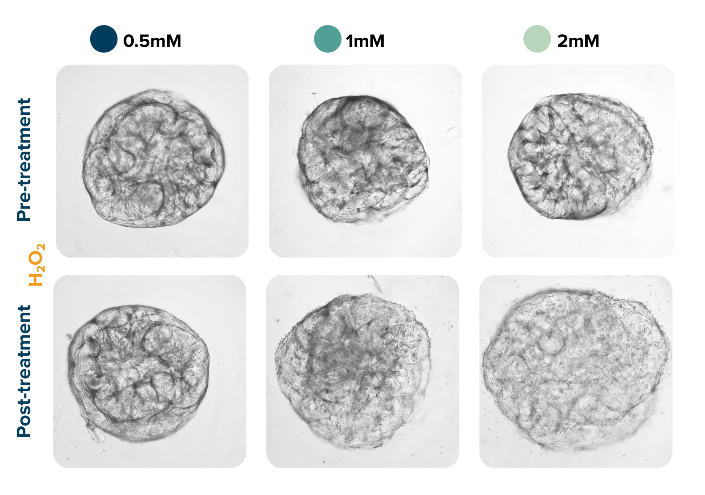
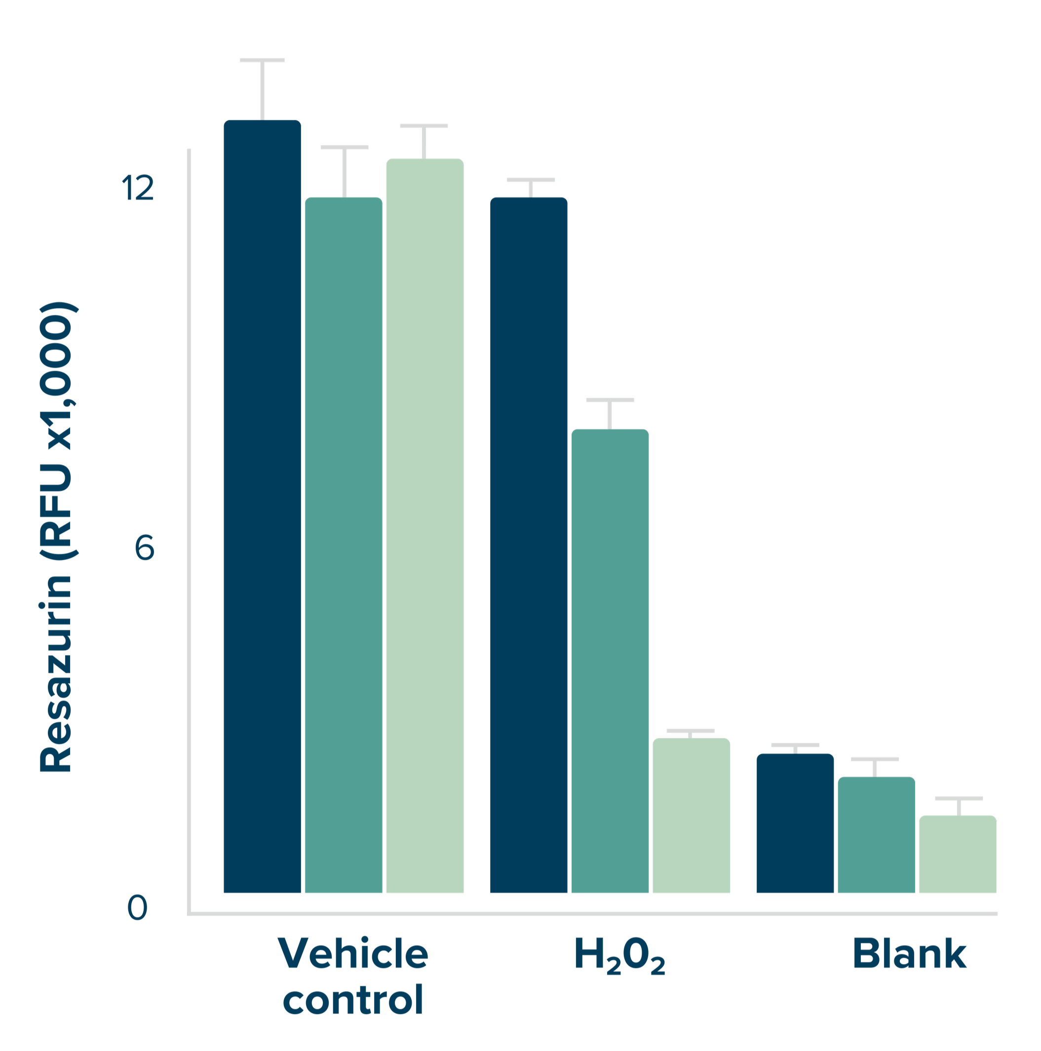
ArtiBrain™ and ADBrain™ provide acute and chronic safety insights, detecting cytotoxicity, oxidative stress, and tissue integrity in 3D neural tissue. These readouts define safe dosing ranges and uncover toxicity before moving into costly animal or clinical studies.
De-risk your compounds with early human-relevant safety data.

Cytotoxicity
Cell Viability
Key assays & readouts:
Cell viability: Resazurin, CellTiter-Glo® 3D
Cytotoxicity: LDH release (luminescence, LDH-Glo®)
Oxidative stress: ROS-Glo® H₂O₂ assay
Imaging: tissue volume and abnormal swelling
Neurodegeneration markers: Tau, pTau, Nf-L
Neuroinflammation
RealBrain® services give you direct access to human-relevant neuroinflammation data. By quantifying glial activation, cytokine release, and inflammatory stress in 3D brain tissue, you gain clear insight into how compounds modulate these pathways — supporting confident, informed development decisions.
From cytokines to cell markers — comprehensive neuroinflammation profiling
In ADBrain™, a small molecule screen quantified amyloid load by imaging, with ELISA we identified that compound B had neuroprotective potential.

Key assays & readouts:
GFAP quantification: imaging & immunoassay
Cytokine & chemokine profiling: single & multiplex immunoassays
Secretome analysis
Imaging: tissue swelling & volumetric changes
Neurodegeneration markers: pTau, Nf-L
Target Validation & Biomarker Discovery
Link compound effects to measurable human biomarkers.
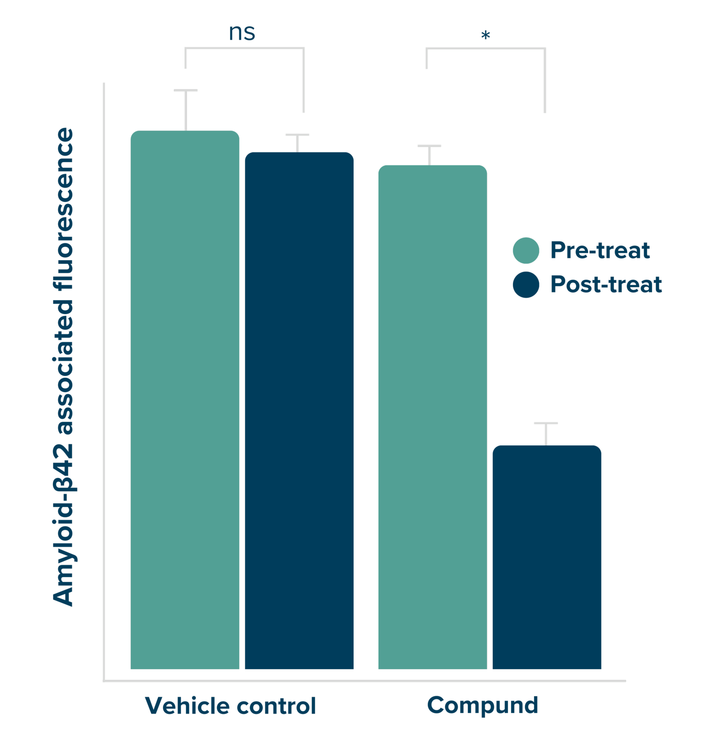
Tessara’s services provide biomarker-driven insights into target engagement and synaptic health, using ArtiBrain™ for baseline and ADBrain™ for pathology to deliver precise, human-relevant molecular data that supports confident development decisions.
In client studies, ADBrain™ showed a 50% reduction in Aβ42 and a 26% increase in neuronal viability, highlighting its value for assessing amyloid-targeting compounds and neuronal health.
Key assays & readouts:
Imaging & immunoassay:
Changes in: Amyloid load (Aβ), total tau, and p-tau variants (p-tau181, p-tau217, p-tau231).
Synaptic plasticity: PSD95 and synapsin
Cellular health: Neurofilament light chain (Nf-L), GFAP, S100B
Omics profiling - genomics & proteomics
Intracellular signaling: pathway activation or inhibition - immunoassay, Western Blot, proteomics
Neurodegeneration & Cell Death
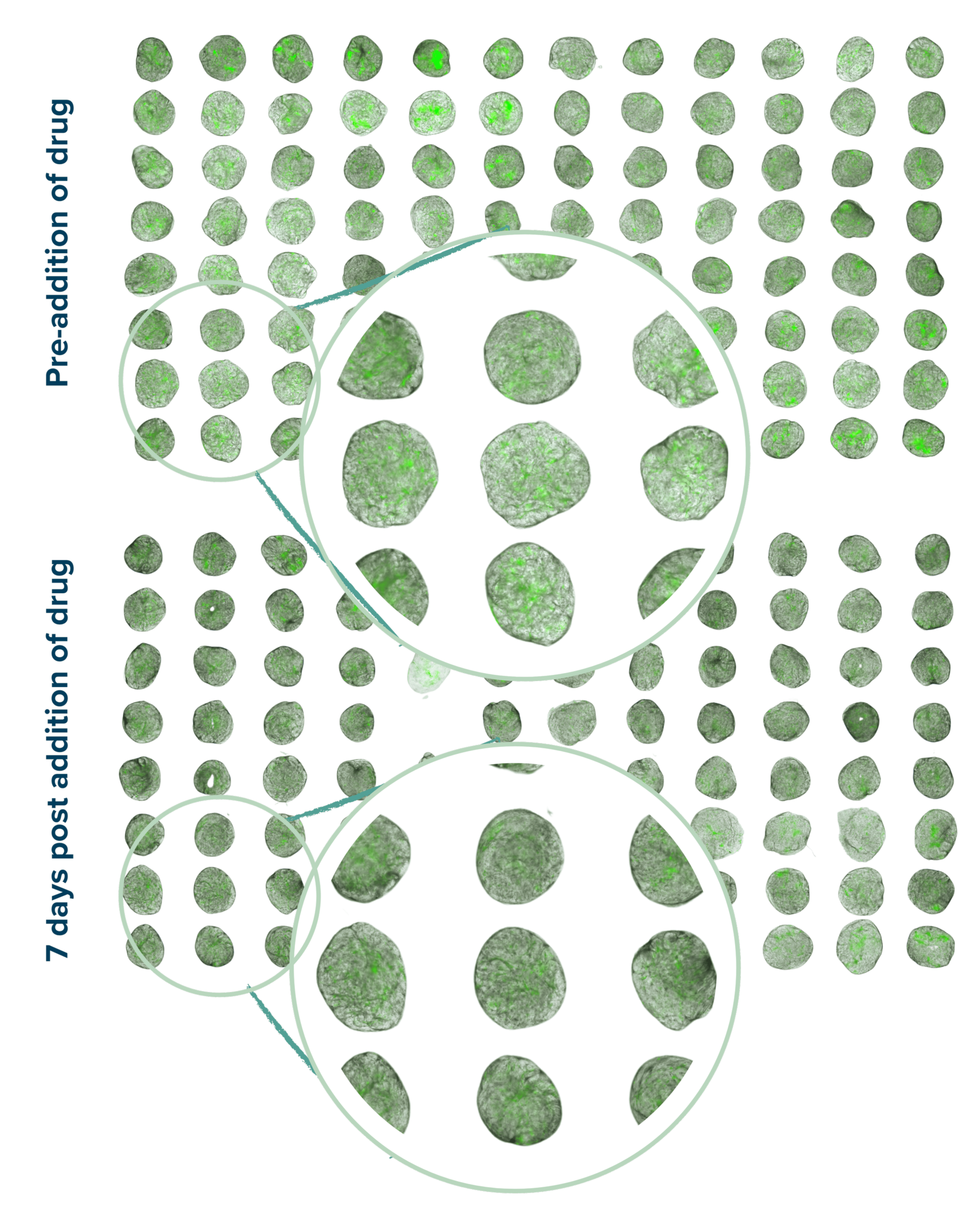
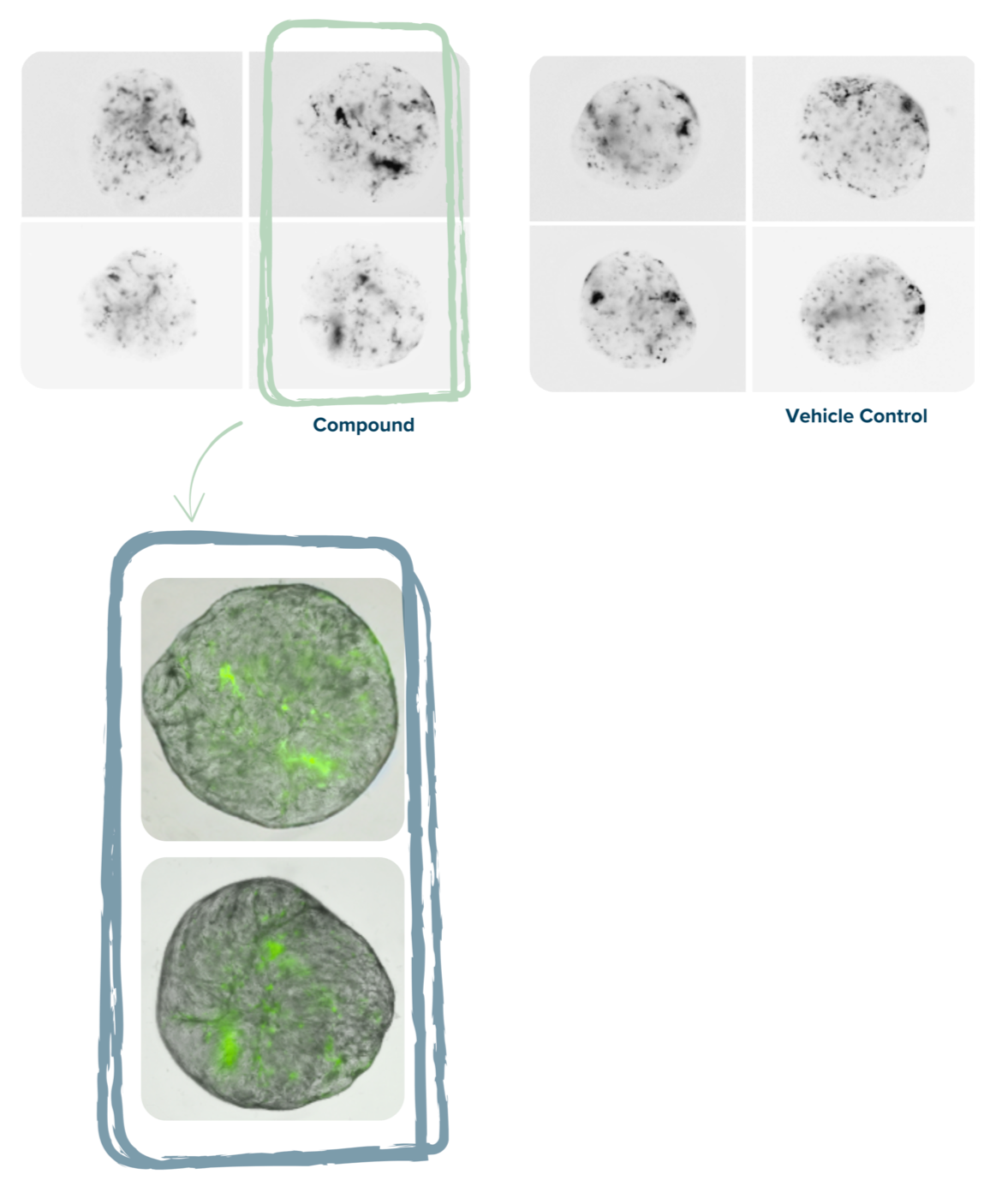
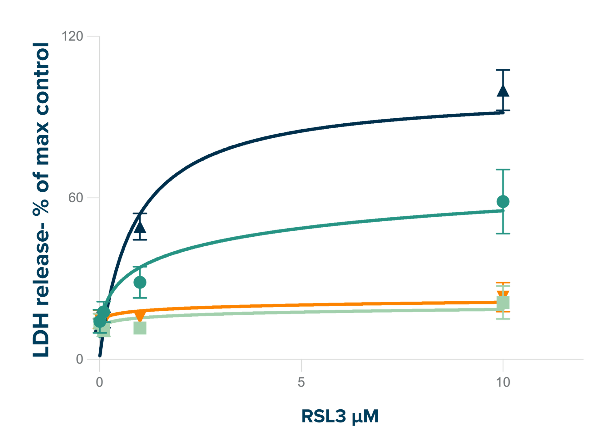
ArtiBrain™ and ADBrain™ capture neurodegenerative pathways, including ferroptosis and neuronal death. These models allow you to compare baseline vs Alzheimer’s sensitivity and test the ability of compounds to prevent or reverse degeneration.
Profile mechanisms of neurodegeneration, with emphasis on neuronal cell death - ferroptosis and injury pathways.

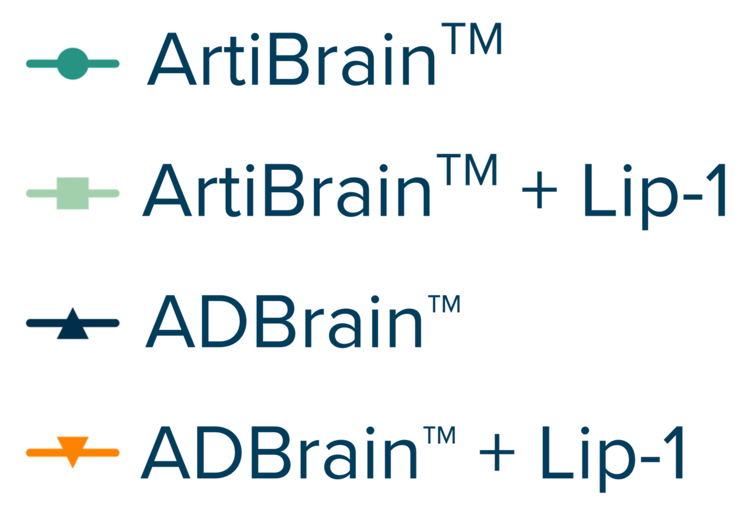
ArtiBrain™ and ADBrain™ respond to RSL3-induced ferroptosis with reduced viability, with ADBrain™ showing higher sensitivity. In both models, Liproxstatin-1 effectively blocked ferroptosis, demonstrating their utility for evaluating compounds in ferroptosis-driven pathology.

Cell Viability
Cytotoxicity
Key assays & readouts:
Cell death pathways: Ferroptosis detection assays
Imaging assays: confocal & brightfield for structural changes
Neurodegeneration markers: pTau, Nf-L
Neural Network Analysis
Measure connectivity, synaptic plasticity, and rescue effects.
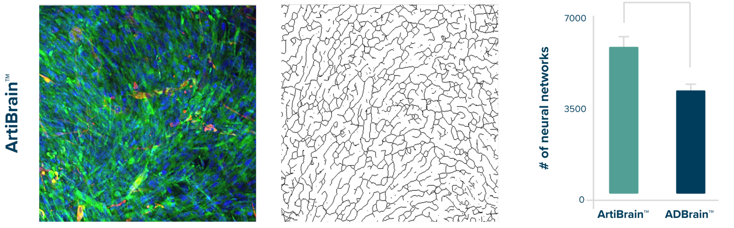
Within three weeks, RealBrain® tissues form dense networks. Using ArtiBrain™ (healthy) and ADBrain™ (disease), we quantify network outgrowth, synaptic changes, and functional recovery following treatment.
Mapped Neural Networks
Neurons Astrocytes

ADBrain™ captures Alzheimer’s-related neurodegeneration, showing reduced networks and shorter neurites. Confocal imaging and automated analysis detect treatment-driven improvements and support biomarker discovery for restoring connectivity.
Key assays & readouts:
Neural outgrowth and branching quantification: Immunocytochemistry and imaging assays - neuronal markers β(III)-tubulin (Tuj1), MAP2, Neurofilament.
Synaptic marker analysis: Immunocytochemistry and imaging assays - PSD95, synapsin
Imaging: High-content confocal imaging and automated image analysis.
Let’s design a study that gives you the answers you need
Turn complex brain biology into clear, decision-ready data — start with RealBrain® services today.